Entamoeba hartmanni is a tiny parasite that lives in the intestines of humans. While often overlooked, it plays an important role in understanding gut health and disease. This post will explore the characteristics of Entamoeba hartmanni, how it differs from similar organisms, and its potential impact on human health. By the end, you will gain valuable insights into this lesser-known protozoan and its significance in the field of microbiology.
Through this article, you’ll be able to answer the following questions:
- What is the difference between Entamoeba histolytica and Entamoeba hartmanni?
- Is Entamoeba hartmanni pathogenic?
- How do you get entamoeba hartmanni?
- What is the habitat of Entamoeba Hartmanni?
Wherever E. histolytica is found, Entamoeba hartmanni is present as well. Until recently, it was thought to be a “small race” of the latter. It was frequently reported as E. histolytica after being misidentified as such. It is currently regarded as a distinct species of commensal intestinal amoeba that is not harmful.
Classification
| Subphylum | CLASS | EXAMPLES OF SPECIES |
| Sarcodina | Lobosea | Entamoeba hartmanni |
Morphology
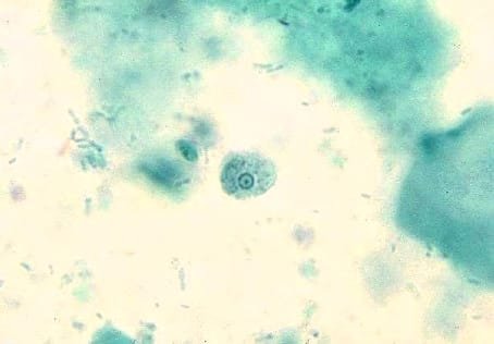
E. hartmanni Trophozoite Characteristics
- Measures 8 to 12 µm, with a size range of 5 to 15 µm.
- Finger-shaped pseudopods with nonprogressive motility.
- Contains one non-visible nucleus.
- Presents peripheral chromatin as evenly distributed granules with a beaded appearance.
- Karyosome may be centrally or eccentrically located.
- Nuclear structure variations similar to E. histolytica.
- Finely granular cytoplasm may contain bacteria.
- Cytoplasm does not contain ingested red blood cells.

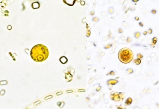
E. hartmanni Cyst Size Overview
- Cysts range from 5 to 12 µm, with an average size of 7 to 9 µm.
- Spherical cysts may have one, two, three, or four nuclei.
- Nuclei in E. hartmanni cysts are smaller than those of its corresponding trophozoite.
- Nuclear structures of fine and peripheral chromatin surrounding a small central karyosome are consistent with E. histolytica.
- Cysts develop similarly to E. histolytica.
- Young cysts have a diffuse glycogen mass and round-ended chromatoid bars.

E. hartmanni Life Cycle
- Similar to other intestinal amoebae.
- Reproduction through binary longitudinal fission.
- Excystation: cyst hatches, converts to trophozoite.
- Binary fission persists, trophozoites reproduce in colon’s lumen.
Disease Transmission
- E. hartmanni Amoebiasis Transmission
- Transmitted through ingestion of food and water.
- Infects cysts in the organism.
- Not considered a pathogen in large numbers.
Clinical Disease (Signs and symptoms)
E. hartmanni: Nonpathogenic Enteric Amoebae
- Typically found in microscopic feces.
- Infections result in asymptomatic condition.
- Often found alongside other pathogenic enteric amoebae.
Diagnosis of E. hartmanni Infection
- Microscopic examination of fresh fecal specimen for ova and parasites.
- Permanent stained smear best for identification.
- Immature and mature cyst forms, trophozoites may be found.
Treatment and Prevention
- Nonpathogenic E. hartmanni Infections
- No treatment needed.
- Cleanliness of food and water is crucial
- Good sanitation and availability of clean water/food eliminate infections.
Conclusion
In summary, understanding E. hartmanni is crucial for effective disease management. Its classification, morphology, lifecycle, and transmission methods highlight the importance of accurate diagnosis and treatment. Recognizing the signs and symptoms early can lead to better health outcomes. Stay informed and take proactive steps in your health journey. For more information on E. hartmanni, consult healthcare professionals or trusted sources. Your health matters, so act wisely.
Bibliography:
- Elizabeth A. Gockel-Blessing (formerly Zeibig), Clinical Parasitology: A PRACTICAL APPROACH. Second Edition. P.184 CHAPTER 7 Miscellaneous Protozoa
- Kayser, Medical Microbiology © 2005 Thieme
- Parasitology for Medical and Clinical Laboratory Professionals by Ridley, John W. Ridley (z-lib.org). Protozoal Microorganisms as Intestinal Parasites 61


